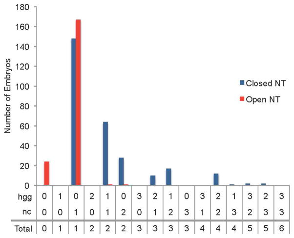
Figure 10. Correlation between total mesendodermal/mesodermal tissue presence and neural tube phenotype.
Combination of data from sqt mutant embryos and embryos exposed to SB505124 starting at 4.3 hpf. Embryos were fixed at 24 hpf and assayed for neural tube phenotype with otx5 expression in the pineal. An elongated or divided pineal indicated an open neural tube while an oval pineal indicated a closed neural tube. The presence of the mesendodermal/mesodermal tissues hatching glands (hgg) and whole anterior-posterior notochord (nc) and were labeled with hgg1 and col2a1 expression, respectively. These tissues were scored for amount present using the following: 3 = full tissue present, 2 = more than half of tissue present, 1 = less than half of tissue present, 0 = tissue absent.

