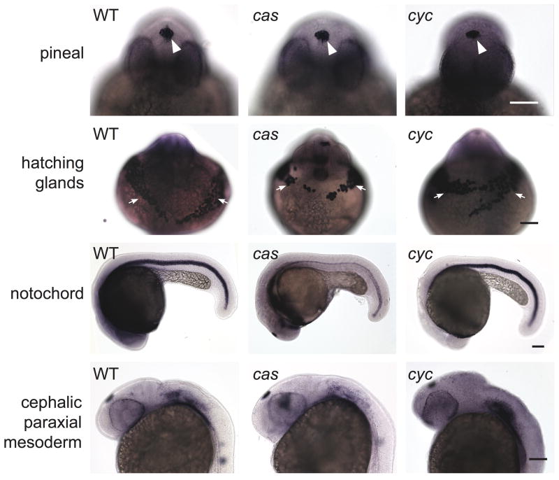
Figure 9. Closed neural tubes and normal mesendoderm/mesoderm development in cyc and cas mutants.
Embryos were co-assayed for otx5 expression in the pineal precursors and a combination of the following mesendodermal/mesodermal markers: ctsl1b in the hatching glands, pax 2.1, col9a2 in cephalic paraxial mesoderm, and shh (for cas mutant) or col2a1 in the notochord (WT embryo and cyc mutant). Embryos in the first three rows are at ~26 somite stage and those in the last two rows are 30 hpf. Images in the same column and of the same age are different views of the same embryo. White arrowheads point to pineal, white arrows to hatching glands. Scale bars: 100 μm.

