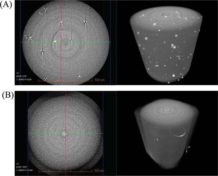
FIGURE 6.

The microscale morphologies of control (A: the left = CT slice at x-y plane from 3D image; the right = 3D image) and experimental (B: the left = CT slice at x-y plane; the right = 3D image) adhesives cured in the presence of 11 wt % water. The morphologies were observed using three-dimensional (3D) MicroXCT (Xradia Inc. Concord, CA). [Color figure can be viewed in the online issue, which is available at wileyonlinelibrary.com.]
