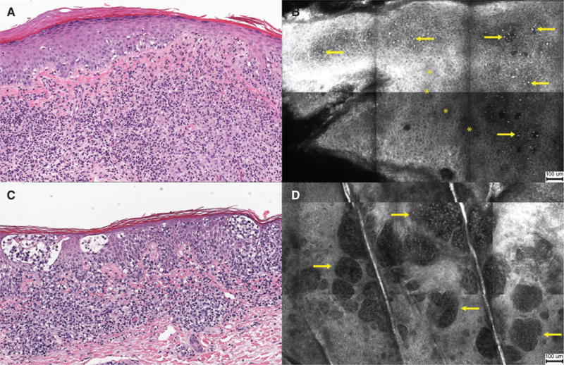
Fig. 3.

Histopathology and reflectance confocal microscopy (RCM) images of plaque lesions. (A) Atypical lymphocytes infiltrating the epidermis [hematoxylin & eosin (H&E) stain, original magnification ×100]. (B) RCM image showing brightly reflective cells (arrows) scattered in the stratum spinosum with focal areas of epidermal disarray (asterisk, *) (scale bar = 100 μm). (C) Collections of atypical lymphocytes forming Pautrier collections within epidermis (H&E, ×100). (D) RCM image showing well-demarcated, vesicle-like, dark spaces containing weakly reflective cells (arrows) (scale bar = 100 μm).
