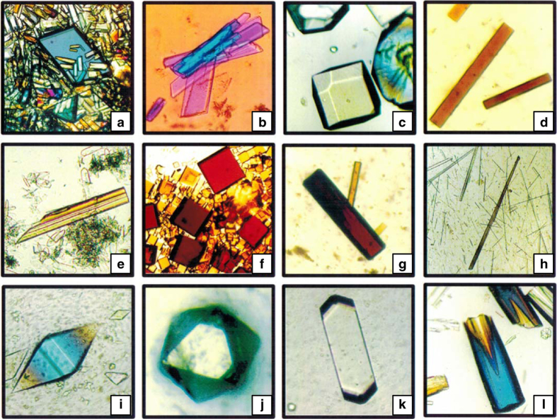
Figure 14.
An array of crystals grown in the dewar device that used liquid–liquid diffusion from frozen biphasic samples. This experiment was performed by American investigators (Koszelak et al.75) on the Russian Space Station Mir. The crystals (labeled by row from left to right) are of top row: (a) rhombohedral canavalin, (b) creatine kinase, (c) lysozyme, (d) beef catalase; middle row: (e) porcine alpha amylase, (f) fungal catalase, (g) myglobin, (h) concanavalin B; and bottom row: (i) thaumatin, (j) apoferritin, (k) satellite tobacco mosaic virus (STMV), (l) hexagonal canavalin.

