. 2017 Jul 18;6:e26591. doi: 10.7554/eLife.26591

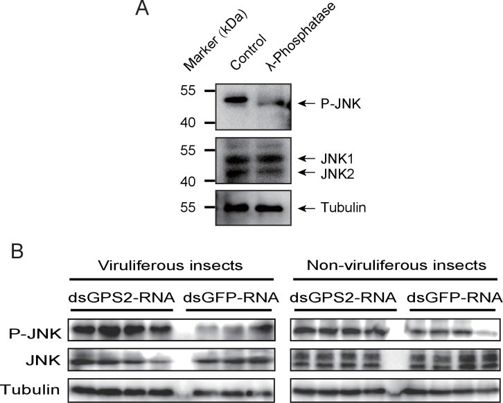
Figure 3. GPS2 represses JNK activation in the planthopper.

(A) Western blot of the phosphorylated JNK (P-JNK) and total JNKs in non-viruliferous fourth instar planthoppers before and after the treatment with λ-phosphatase. Total protein was incubated with λ-phosphatase for 1 hr at 30°C. Three independent biological replicates were carried out. Here we show one representative result. (B) Western blotting showing P-JNK in viruliferous or non-viruliferous fourth instar nymphs when dsGPS2-RNA was injected. Levels were determined 3 d after injections. dsGFP–RNA injection was used as control. The P-JNK, total JNKs, and tubulin were detected using an anti-phospho-human JNK2 antibody, an anti-human JNK2 polyclonal antibody, and an anti-human tubulin monoclonal antibody, respectively.

DOI: http://dx.doi.org/10.7554/eLife.26591.009

Figure 3.

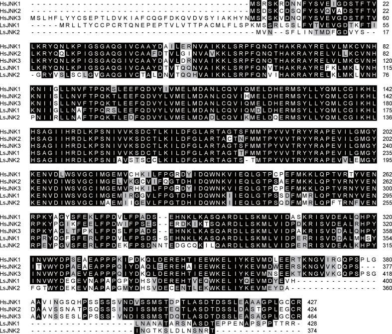
Figure 3—figure supplement 1. Amino acid sequence alignments of planthopper JNKs (LsJNK1 and LsJNK2) and human JNKs (HsJNK1, HsJNK2, HsJNK3).

Figure 3—figure supplement 1.

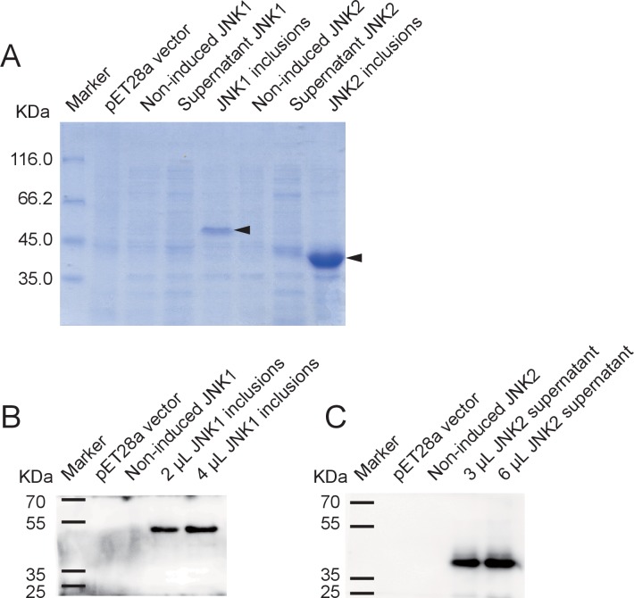
Figure 3—figure supplement 2. Recombinant expression of planthopper JNK1 and JNK2 for specificity verification of the anti-human JNK2 polyclonal antibody.

Figure 3—figure supplement 2.

(A) SDS-PAGE showed the recombinant expression of JNK1 and JNK2 in E. coli. Arrows indicate the target proteins. (B) and (C) Western blot analysis of JNK1 and JNK2 using the anti-human JNK2 polyclonal antibody. JNK1 inclusions and supernatant JNK2 were applied. The 51-kD recombinant JNK1 and 44-kD recombinant JNK2 were revealed.
Figure 3—figure supplement 3. Densitometry analysis for the phosphorylated JNK (P-JNK) image bands from Figure 3B.

Figure 3—figure supplement 3.

The relative densities of P-JNK were normalized with those of tubulin and presented as mean ± SE. *p<0.05.