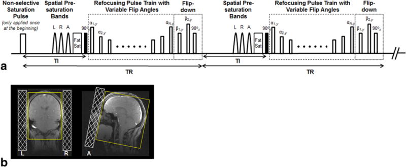
FIG. 1.

(a) Pulse sequence diagram: nonselective saturation pulse is applied only at the beginning to achieve quick transition of magnetizations to a steady state. Three spatial presaturation bands placed on the left (L), right (R), and anterior (A) to saturate the signals from the ears and nose are followed by fat saturation (Fat Sat), nonselective excitation, and a variable-flip-angle refocusing pulse train. A flip-down module is applied immediately after the refocusing pulse train, allowing the remaining magnetizations to be transferred to the z-axis, where all tissues subsequently start to undergo inversion recovery. (b) Schematic demonstration of the location of the L, R, and A saturation bands on localization images. These bands help effectively reduce the required phase and partition-encoding steps, and thus minimize scan times.
