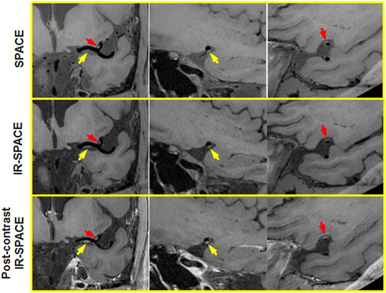
FIG. 3.

A 50-year-old male volunteer with incidental findings. Both SPACE and IR-SPACE in the precontrast state exhibited wall thickening with relatively high signal at the left MCA M1 and M2. Eccentric postcontrast enhancement was also observed at the same locations, indicating atherosclerotic lesions with inflammation. Left column: the in-plane view demonstrating the two suspicious locations (arrows) at MCA M1 and M2, respectively; middle column: the cross-sectional view of the suspicious location at the MCA M1; right column: the cross-sectional view of the suspicious location at the MCA M2.
