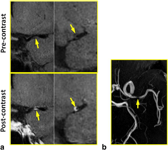
FIG. 5.

In a 57-year-old female patient with atherosclerosis disease, a severe stenosis with eccentric wall thickening and contrast enhancement was detected at MCA by IR-SPACE. Compared with TOF MR angiography, the vessel wall imaging sequence provided more definitive proof that atherosclerosis is involved in the particular part of the vessel wall. (a) Left column: in-plane view demonstrating the stenotic lumen (arrows); right column: cross-sectional view of the atherosclerotic lesion. (b) TOF thin maximum-intensity-projection images confirming the severe stenosis at the same location.
