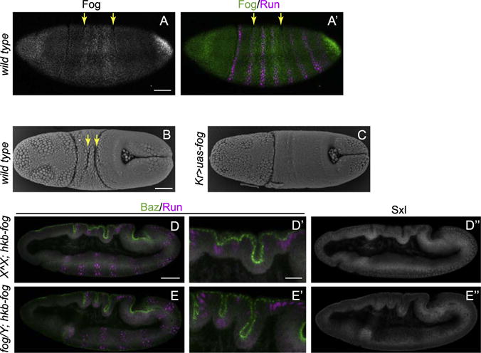
Fig. 6. Fog expression between dorsal folds optimizes the morphology of the epithelial folds.

(A) Fog is expressed at low levels between the future dorsal folds, whose positions are indicated by the second and fifth Runt stripes (yellow arrows). (B–C) Scanning EM images show two transverse furrows (yellow arrows) form in the wild type embryo (B) but are abolished in the embryos with Fog expressed throughout the dorsal ectoderm (C). (D–F) Dorsal folds are deeper in the fog mutant embryos carrying hkb-fog compared to the embryos expressing hkb-fog alone. Male embryos carrying a fog mutant X chromosome and a Y chromosome are identified by the absence of nuclear Sex-lethal (Sxl) staining. Scale bars: 50 μm (A–C, D–E, D″–E″) and 20 μm (D′–E′).
