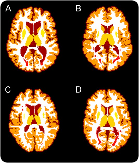
Figure 1. LesionTOADS thalamic segmentation depicted in one participant from each cohort.

(A) MS without comorbidities for MRI white matter abnormalities, (B) MS with an additional comorbidity for MRI white matter abnormalities, (C) migraine with MRI white matter abnormalities and without additional comorbidities for MRI white matter abnormalities, (D) previously misdiagnosed with MS. Dark red: CSF, dark orange: cortical gray matter, light orange: thalamus and striatum, white: white matter, and red: lesions.
