
Figure 2. Thalamic volume normalized to intracranial volume for each participant in the 4 cohorts.
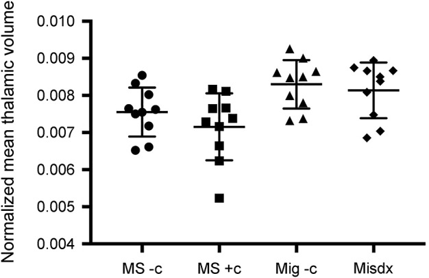
MS − c: MS without comorbidities for MRI white matter abnormalities, MS + c: MS with additional comorbidities for MRI white matter abnormalities, Mig − c: migraine with MRI white matter abnormalities and without additional comorbidities for MRI white matter abnormalities, Misdx: previously misdiagnosed with MS.