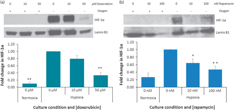
Fig. 3.

Nuclear accumulation of hypoxia-inducible factor 1α (HIF-1α) after doxorubicin and rapamycin treatments. Normoxic and hypoxic HepG2 cells were exposed to doxorubicin or rapamycin for 24 h. Nuclear extracts were fractionated on a 10% SDS-PAGE gel, transferred to a PVDF membrane and probed with anti-HIF-1α antibodies. Proteins were visualized using chemiluminescence. The membrane was stripped and reprobed using antibodies against the nuclear house-keeping protein Lamin B1. Protein levels were quantified using densitometry analysis. HIF-1α was normalized to Lamin. Fold change compared with untreated hypoxic cells was calculated. Statistical analysis was carried out using a t-test. *P<0.05, **P<0.01.
