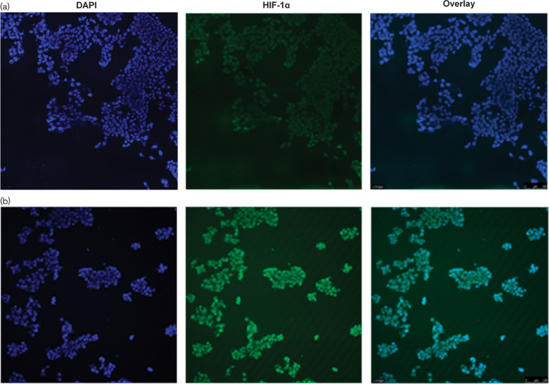
Fig. 4.

Immunohistochemistry staining for hypoxia-inducible factor 1 (HIF-1) in normoxic and hypoxic HepG2 cells. Cells were seeded onto chamber slides. HepG2 cells were seeded onto chamber slides and incubated under normoxic conditions until confluence was 60%. The cells were then incubated under either (a) normoxic conditions or (b) hypoxic conditions for 24 h. The cells were incubated overnight with antibodies to HIF-1α and then incubated with the TRITC-conjugated secondary antibody. 4′,6-Diamidino-2-phenylindole staining was used to visualize the nucleus. Images are taken at ×100 magnification. HIF-1α is absent in normoxic cells and present in hypoxic cells.
