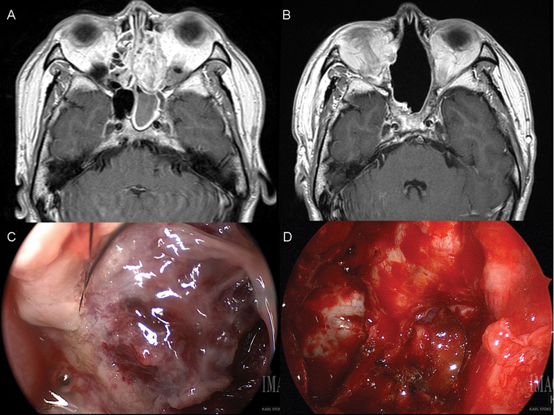
Fig. 1.

( A ) Preoperative and ( B ) 24-month postoperative axial T1-weighted MRI with contrast of the index teratocarcinosarcoma. Intraoperative appearance of sinonasal teratocarcinosarcoma ( C ) before and ( D ) after resection. MRI, magnetic resonance imaging.
