
Figure 6.
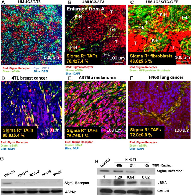
Sigma receptor is expressed in αSMA positive TAFs. (A) IF staining of paraffin embedded tumor sections from the UMUC3/3T3 model. Red: Sigma R; green: αSMA; and cyan: CD31. Co-localization was observed partially between Sigma R and αSMA and illustrated as orange or yellow. (B) Higher magnification of the UMUC3/3T3 model stained with Sigma R and αSMA. The Sigma R high and low regions in fibroblasts were marked as F–H and F–L. Tumor nest (T) was highly positive for Sigma R. (C) Cryosections of UMUC3/3T3. Green indicated GFP-positive fibroblasts, and red indicated Sigma R. (D–F) Costaining of Sigma R (red) and αSMA (green) in 4T1, A375lu, and H460. The % of Sigma R positive fibroblasts or TAFs (αSMA) were quantified by ImageJ (n = 3). (G,H) Western blot analysis of Sigma R expression in vitro on different cell lines (including tumor cells UMUC3, and other mouse or human fibroblasts). Note that activation of NIH3T3 with TGFβ enhances the expression of Sigma R. The numbers below Western blot of Sigma R indicate the average intensity (quantified by ImageJ) of the blot in each samples compared to UMUC3 (set as 1).