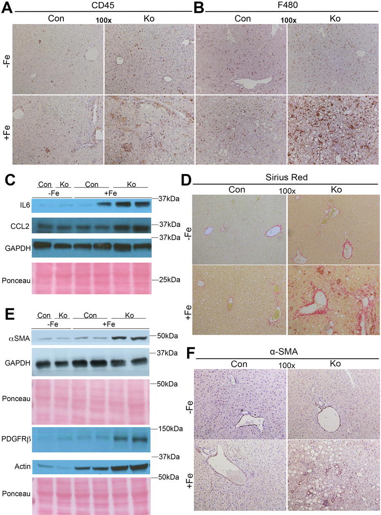
Figure 2. KO+Fe have increased hepatic inflammation and fibrosis after chronic iron overload.

(A) Representative IHC images demonstrate increased CD45-positive inflammatory cells in KO+Fe as compared to other groups. (B) Representative IHC images demonstrate increased F480-positive macrophages in KO+Fe only. (C) Representative WB shows KO+Fe have increased inflammatory markers including IL-6 and CCl2. IL-6 and CCl2 appear to be migrating slower due to glycosylation. (D) KO+Fe have increased Sirius Red staining suggesting increased collagen deposition. (E). WB show increased levels of α-SMA and PDGFRβ substantiating presence of fibrosis. (F) IHC for α-SMA also confirms presence of activated myofibroblasts in KO+Fe only. (Image magnification: 100×)
