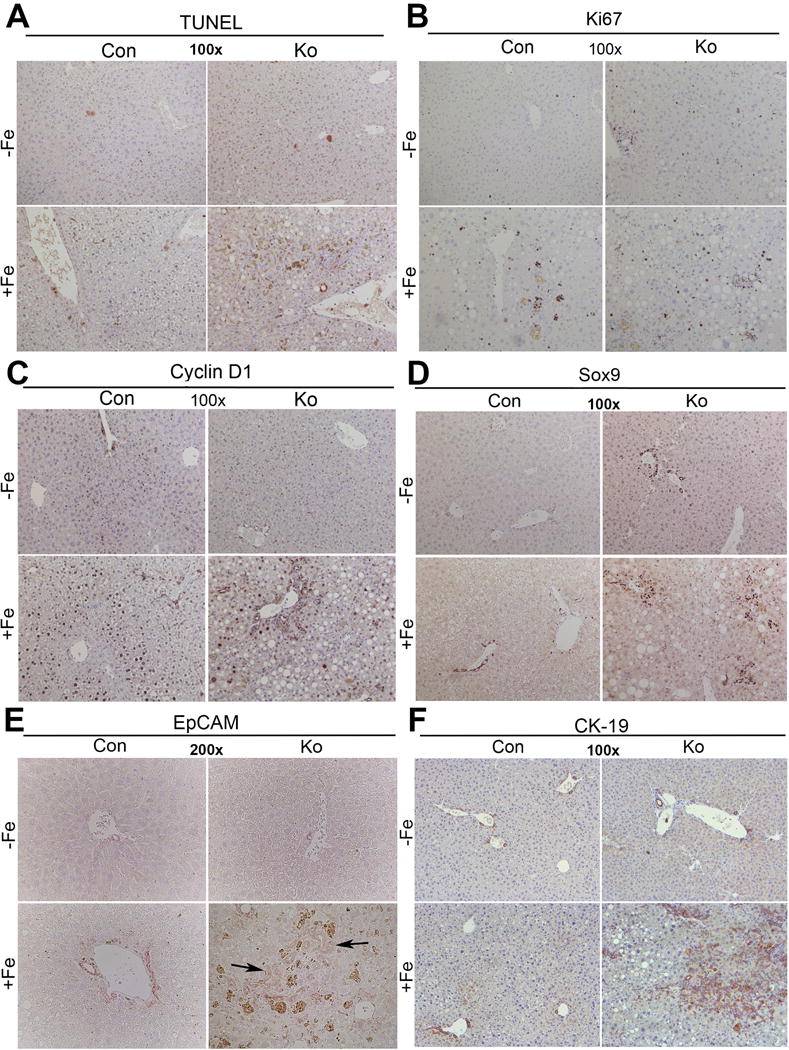
Figure 3. Analysis of cell death, proliferation and ductular reaction in CON and KO on normal and high iron diet.

(A) Representative images of TUNEL staining shows marginal increase in cell death in CON+Fe, but a greater increase in KO+Fe. (B) Ki-67 IHC demonstrates comparable and increased number of cells in S-phase after iron overload in both CON+Fe and KO+Fe. (C) Cyclin-D1 staining is increased in midzonal hepatocytes in CON+Fe compared to CON−Fe. KO+Fe exhibit less Cyclin-D1 staining in periportal hepatocytes and in ductular cells. (D) KO+Fe have increased ductular reaction as evident by IHC for Sox9. (E) Increased ductular reaction in KO+Fe was confirmed by EpCAM staining. (F) Increased ductular reaction in KO+Fe was also confirmed by IHC for CK19. (Image magnification:100×, except EpCAM:200×).
