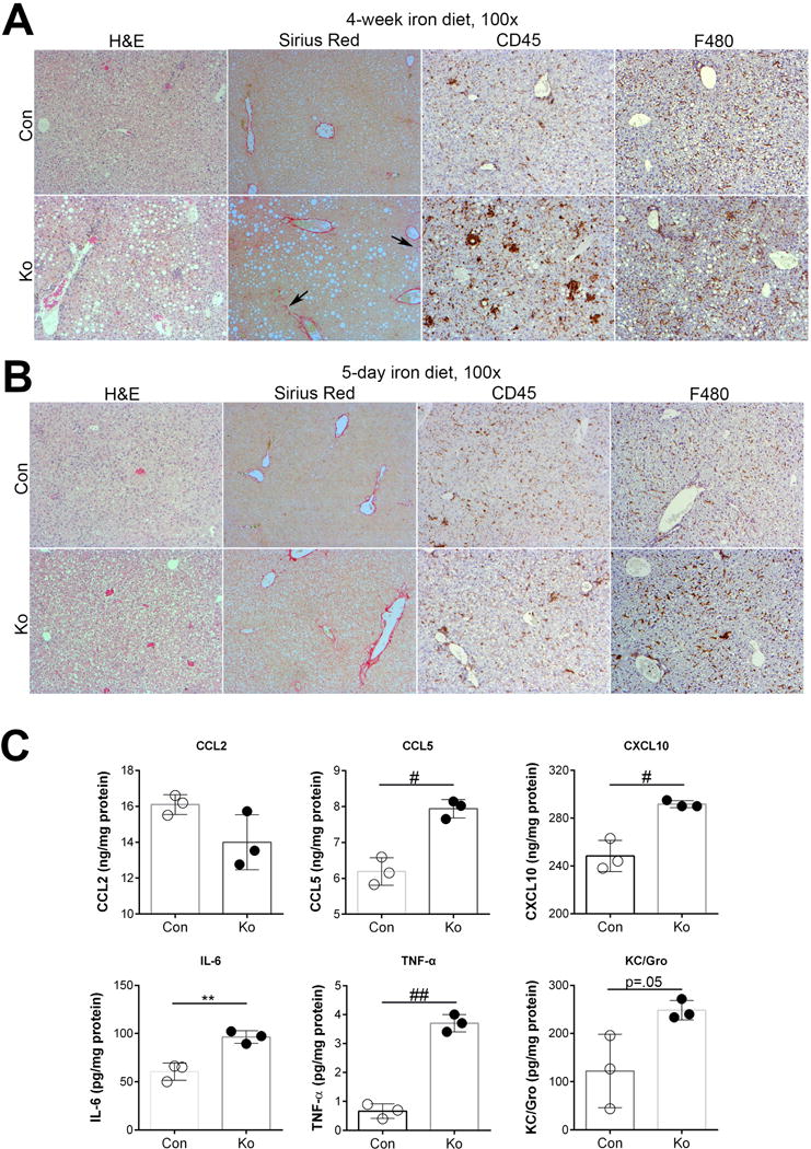
Figure 4. Increased levels of inflammatory cytokines precede steatohepatitis and fibrosis in KO+Fe as shown by time course after iron-feeding.

(A) H&E, Sirius Red and IHC for CD45 and F480 in CON and KO livers after 4-week iron diet shows presence of steatohepatitis in KO+Fe and initiation of fibrosis. (B) H&E, Sirius Red and IHC for CD45 and F480 in CON and KO livers after 5-day iron diet. Con+FE and KO+Fe lack steatosis and fibrosis while CD45-positive inflammatory cells and F480-macrophages appear to be comparably increased in both groups. (C) Cytokine assay using liver lysates from 5-day CON+Fe and KO+Fe reveals comparable increased protein levels of CCL5, CXCL10, IL-6, TNFα, and KC/Gro in KO+Fe but no change in CCl2. *p<0.05, **p<0.01, #p<0.005, #p<0.001 using Student’s T test. (Image magnification: 100×)
