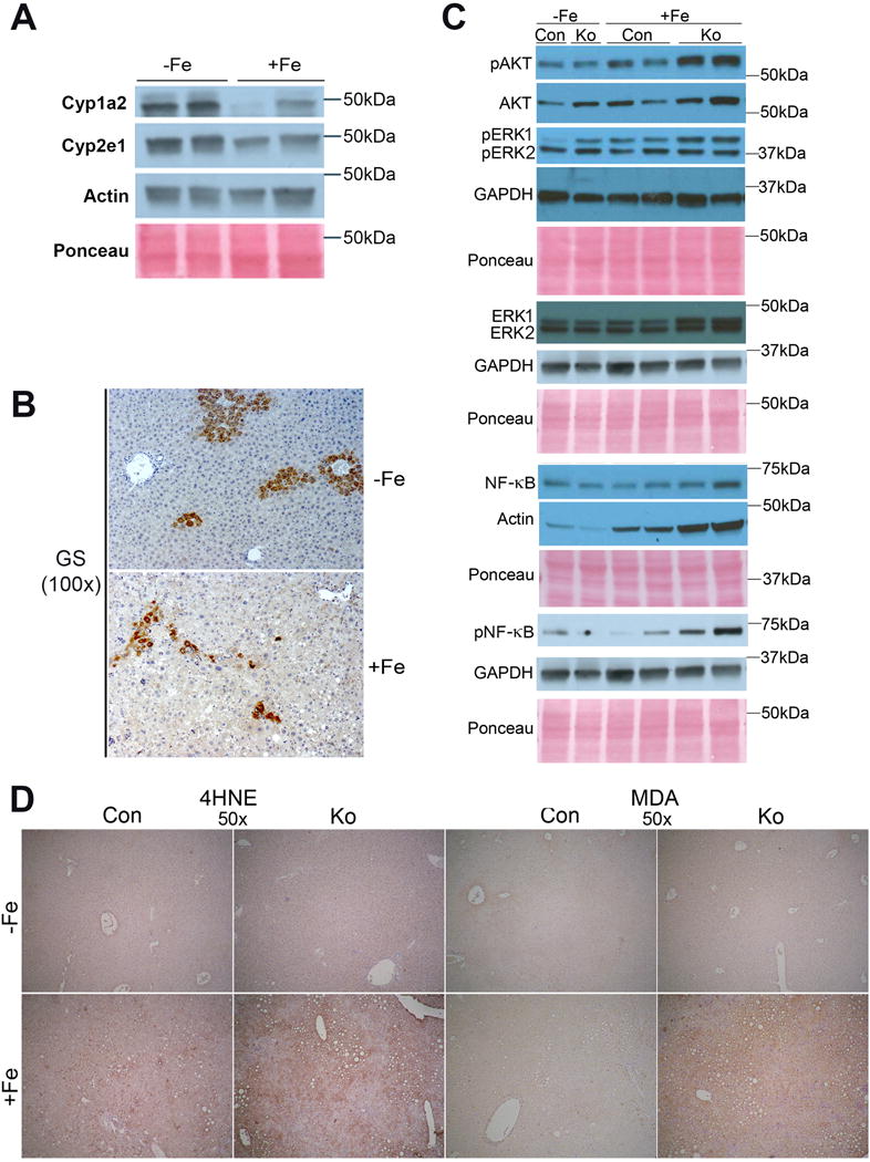
Figure 6. Chronic iron overload affects β-catenin signaling in CON+Fe, but induces AKT, ERK, and NFκB signaling and lipid peroxidation in KO+Fe.

(A) Representative WB show a decrease in hepatic levels of Cyp1a2 and Cyp2e1 levels after iron overload. (B) Representative IHC for GS shows decreased uniformity of GS staining in pericentral hepatocytes in CON after iron overload (100×). (C) Representative WB show greater increases in pAKT (Thr308) and pERK1 (Thr202/Thr204) in KO+Fe as compared to CON+Fe. pNFкB (Ser536) is induced only in KO+Fe. (D) Representative IHC images show increased staining for lipid peroxidation markers 4-hydroxynonenal (4HNE) and malondialdehyde (MDA) in KO+Fe. (50×).
