
Figure 2.
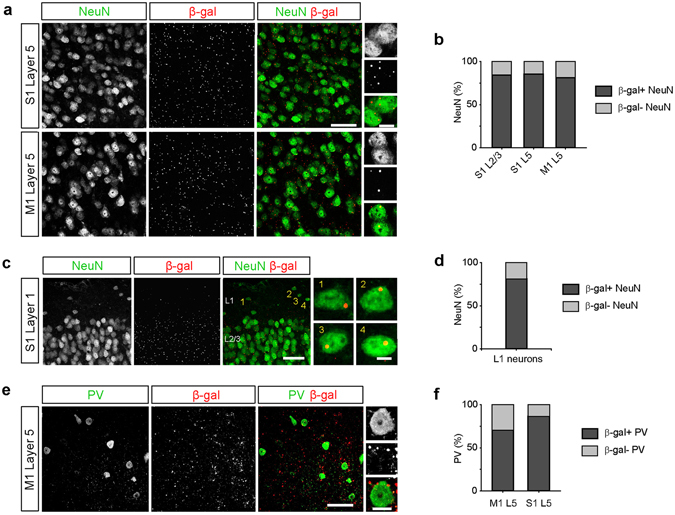
C9orf72 promoter activity is widespread in inhibitory neurons. (a) Representative images of layer 5 (L5) of primary somatosensory cortex (S1, top) and primary motor cortex (M1, bottom) from C9orf72 LacZ/+ mice stained for NeuN (green) and β-galactosidase (β-gal; red). High magnification images show β-gal-positive puncta within NeuN-positive neurons (rightmost panels). Scale bars: 50 μm and 10 μm. (b) The percentage of neurons containing β-gal puncta in layer 2/3 and L5 of somatosensory cortex and L5 of motor cortex (S1 L2/3: 362 of 429 cells, 84.4%; S1 L5, 499 of 583 cells, 85.6%; M1 L5: 425 of 523 cells, 81.3%; n = 3 mice for each group, p = 0.1378, Chi-Square test). (c) Representative images of layer 1 (L1) of somatosensory cortex stained for NeuN (green) and β-gal (red). High magnification images showing β-gal-positive puncta within L1 neurons (rightmost panels). Scale bars: 50 μm and 5 μm. (d) The percentage of L1 neurons containing β-gal puncta (26 of 32 cells, 81.3%, n = 3 mice). (e) Representative images of L5 of motor cortex stained for parvalbumin (PV, green) and β-gal (red). High magnification images show β-gal puncta within a PV interneuron (rightmost panels). Scale bars: 50 μm and 10 μm. (f) The percentage of PV interneurons containing β-gal puncta in L5 of motor cortex and somatosensory cortex (M1 L5: 36 of 51 cells, 70.6%; S1 L5: 32 of 37 cells, 86.5%; n = 3 mice for each group, p = 0.0789, Chi-Square test).