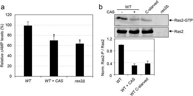
Figure 5.

Intracellular cAMP and Ras2-GTP levels decrease in cells treated with caspofungin. (a) Wild-type (WT) was grown in YPD medium in the absence or presence of CAS during 2 h for cAMP quantification. The ras2Δ mutant was included as control. Relative cAMP levels are shown (wild-type strain = 100%) from three independent experiments. Statistical analysis was carried out using a two-tailed, unpaired, Student’s t-test to analyse differences between the wild-type strain treated with CAS or the ras2∆ mutant versus the wild-type strain without treatment: 0.01 ≤ *p ≤ 0.02. (b) Whole cell extracts were prepared from early log phase cells of the wild-type (WT) strain grown in YPD in the presence or absence of CAS for 2 h, the ras2Δ strain grown in YPD and WT cells grown in SC medium transferred to SC-D for 2 h (conditions of glucose starvation, C-starved). Active Ras2 (Ras2-GTP) was pulled down using GST-RBD bound to glutathione beads. The levels of Ras2-GTP (in the pull-down samples) as well as total Ras2 (in the whole cell extracts) were detected by immunoblotting with the anti-Ras2 antibody. A representative Western blot is shown. The mean and SD from three independent experiments of the ratio Ras2-GTP/total Ras2 relative to the wild-type sample (ratio = 1) is presented in the lower panel.
