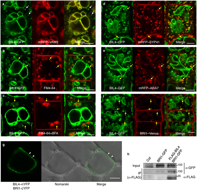
Figure 5.

BIL4 is localized to punctate structures and the vacuolar membrane and interacts with BRI1. (a) BIL4pro::BIL4–GFP partially co-localizes with the vacuolar membrane marker mRFP–VAM3 (unmerged puncta are marked by white arrows). (b) BIL4pro::BIL4–GFP partially colocalizes with FM4-64. Seedlings were treated with FM4-64 for 5 min and then incubated in water for 40 min. (c) Seedlings were pretreated with FM4-64 for 5 min, incubated in water for 20 min and then treated with 50 μM BFA for 20 min. (d–f) BIL4pro::BIL4–GFP partially co-localizes with the TGN/EE marker mRFP–SYP41 (d), the LE/MVB marker mRFP–ARA7 (e) and BRI1pro::BRI1–Venus in the endosome (f). Merged structures in b–f are marked by yellow arrows. Scale bar in (a–f): 5 µm. (g) BiFC assay of the interactions between BIL4 and BRI1 in cultured Arabidopsis cells. Scale bars, 10 μm. The white arrowheads indicate characteristic interactions. (h) Co-immunoprecipitation of BRI1 and BIL4. Wild-type (Col-0) and transgenic plants coexpressing BRI1pro::BRI1–GFP and 35 S::FLAG–BIL4 were grown for 7 days. FLAG–BIL4 was immunoprecipitated by anti-FLAG antibody, and the immunoblots were probed with anti-GFP or anti-FLAG antibody.
