Figure 6.

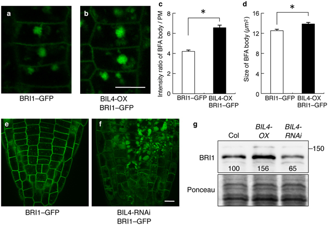
BRI1 subcellular localization is affected in the BIL4-OX and BIL4-RNAi plants. (a–d) Three-day-old seedlings were treated with BFA (50 μM, 0.5 hr). BRI1–GFP-labeled BFA bodies in the wild-type (a) and BIL4-OX plants (b). Scale bar, 10 µm. Signal intensities of BRI1–GFP-labeled BFA bodies in the wild-type and BIL4-OX plants (c). Sizes of BRI1–GFP-labeled BFA bodies in the wild-type and BIL4-OX plants (d). (c,d) n = 3 roots with at least 30 BFA bodies. *P < 0.01, Student’s t-test. Mean ± s.e. (e,f) BRI1–GFP localization in the root tip of wild-type (e) and BIL4-RNAi mutant (f) 2 days after germination in the dark. Scale bar, 10 µm. (g) Plants were grown in the dark for 7 days on medium containing 3 µM Brz. Western blot analyses were performed using the anti-BRI1 antibody (upper panel). The protein levels were detected using Ponceau S (lower panel). Numbers indicate the relative BRI1 signal levels normalized to the Ponceau S-stained protein band.
