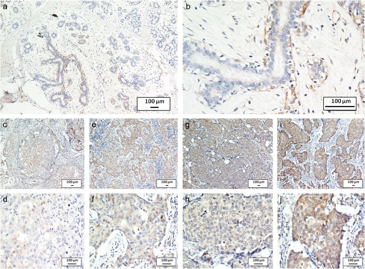
Fig. 2.
CST1 expression analyzed by immunohistochemical staining. Negative CST1 staining in normal breast ductal epithelium (negative control) a ×100, b ×400, negative staining of CST1 in breast cancer tissue c ×100, d ×400, weak staining of CST1 in cytoplasm e ×100, f ×400, moderate staining of CST1 in cytoplasm g ×100, h ×400, and strong staining of CST1 in cytoplasm i ×100, j ×400

