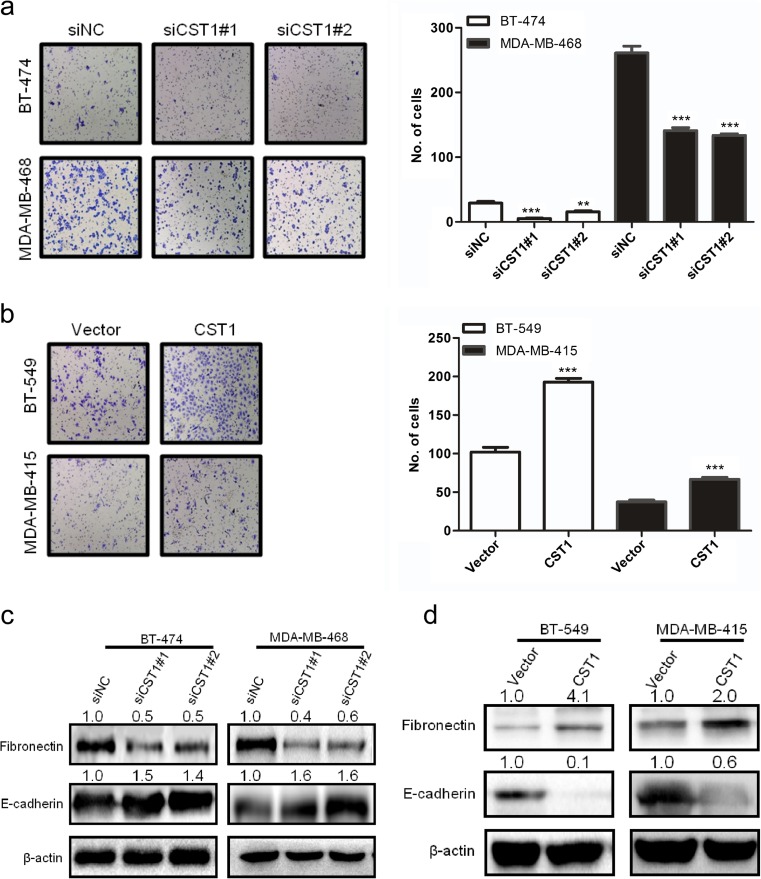
Fig. 8.
CST1 promotes migration in breast cancer cells. a The representative pictures and quantification of migration in BT-474 and MDA-MB-468 cells transfected with NC- or CST1-targeting (KD) siRNAs by transwell migration assay after the knockdown of CST1. b The representative pictures and quantification of cell migration in BT-549 and MDA-MB-415 cells harboring vector or pMSCV-CST1 plasmid by transwell migration assay. c The representative pictures of metastasis-related proteins after the knockdown of CST1, as determined by western blotting. d The representative pictures of metastasis-related proteins after the overexpression of CST1, as determined by western blotting. Migrating cells were identified using light microscope (×100). Results are expressed as means ± SD (error bars) from five viewing fields. *p < 0.05, **p < 0.01, and ***p < 0.001 compared to siNC or vector

