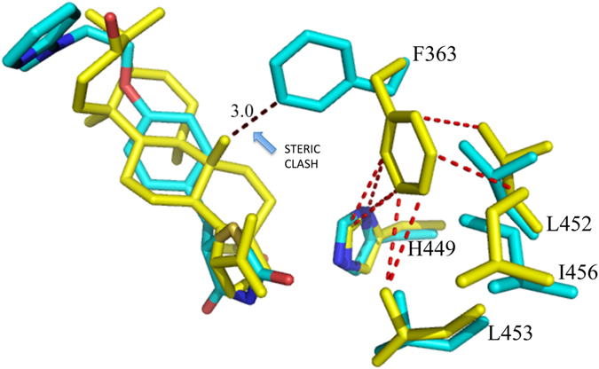
Figure 4.

Superposition of PPARγ/BA and PPARγ/rosiglitazone complexes. Superposition of the crystal structures of the PPARγ/BA (yellow) and PPARγ/rosiglitazone (pdb code 2PRG) (cyan) complexes. The F363 side-chain in gauche conformation (cyan) would sterically clash (black dashed lines) with a methyl group of BA. The gauche* conformation of F363 allows vdW interactions (ranging from 3.3 to 3.7 Å) with hydrophobic residues on H11 (red dashed lines).
