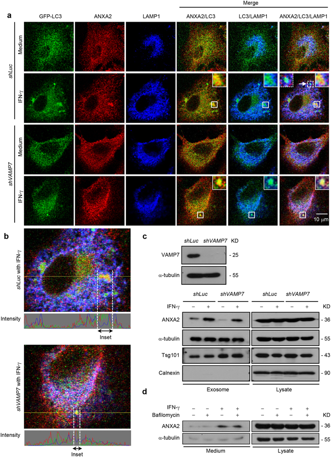
Figure 4.

Amphisome/lysosome fusion is not required for autophagy-mediated exosomal secretion of ANXA2. (a) Cells with VAMP7 knockdown and control knockdown were transfected with a GFP-LC3 plasmid and treated with or without 500 U/ml IFN-γ for 24 h. Cells were then fixed, permeabilized, and stained for ANXA2 (red) and LAMP1 (blue). The colocalization of ANXA2, GFP-LC3 and LAMP1 was observed by confocal microscopy. Scale bar: 10 μm. The arrow and dotted inset mark an autolysosome. (b) Line tracing analysis of fluorescence signal from image in (a) of VAMP7 knockdown and control knockdown cells after IFN-γ stimulation is shown. (c) VAMP7 knockdown efficiency was detected by western blotting. Control and VAMP7-silenced cells were treated with or without 500 U/ml IFN-γ for 48 h. The exosome pellets were collected. ANXA2, α-tubulin, Tsg101 and calnexin from exosome pellets and total cell lysates were detected by western blotting. kD, molecular weight as kDa. (d) A549 cells were incubated with 500 U/ml IFN-γ in the presence or absence of 5 nM bafilomycin A1 for 48 h. ANXA2 and α-tubulin from cultured supernatant and total cell lysate were analyzed by western blotting. kD, molecular weight as kDa.
