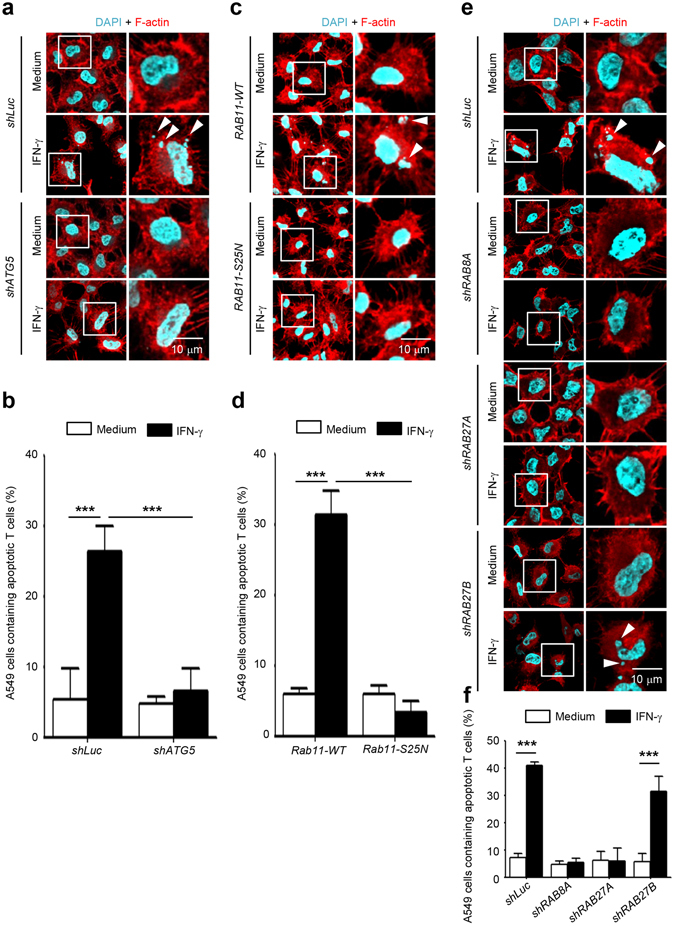
Figure 6.

Inhibition of exophagy of ANXA2 reduces ANXA2-mediated efferocytosis. Jurkat T cells were treated with 50 μM etoposide for 24 h to induce cell apoptosis. ATG5, RAB8A, RAB27A, or RAB27B knockdown or pcDNA-RAB11wt- and pcDNA-RAB11S25N-overexpressing A549 cells were treated with 500 U/ml IFN-γ for 48 h. After treatment, these A549 cells were co-cultured with apoptotic Jurkat T cells for 2 h. Cells were then washed, fixed, permeabilized and stained with DAPI and rhodamine-phalloidin for nucleus and F-actin, respectively. Cells were mounted and analyzed by confocal microscopy. The nuclei of apoptotic T cells were indicated by white arrowheads in ATG5 knockdown cells (a), pcDNA-RAB11wt- and pcDNA-RAB11S25N-overexpressing cells (c), and RAB8A, RAB27A, RAB27B knockdown cells (e). Scale bar: 10 μm. Cells containing nuclei fragments were quantified in ATG5 knockdown cells (b), pcDNA-RAB11wt- and pcDNA-RAB11S25N-overexpressing cells (d), and RAB8A, RAB27A, RAB27B knockdown cells (f). Data are represented as mean ± SD. ***P < 0.001. At least 70 cells were counted in each treatment group.
