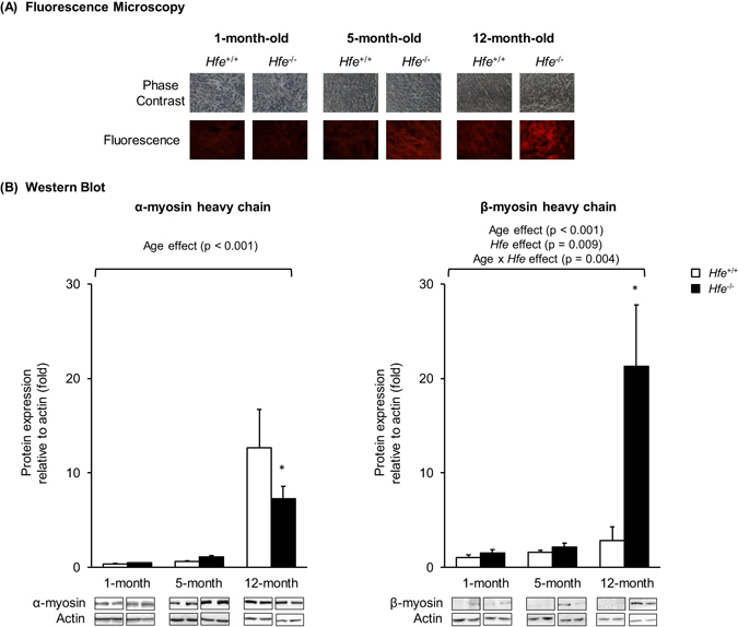
Figure 2.

Effect of age on the expression of cardiac myosin heavy chains in Hfe +/+ and Hfe −/− mice. (A) Cardiac myosin expression in 1-month, 5-month and 12-month old Hfe +/+ and Hfe −/− mice (n = 3–5 per group) was evaluated by fluorescence microscopy. Frozen heart tissues were sectioned using cryostat (10 µm thickness) and stained with bispecific myosin antibody polymer, which was conjugated to anti-dithiopropionic acid-rhodamine isothiocyanate. Sections stained without bispecific polymer were used as background control. (B) Representative immunoblots are shown for α- and β-myosin heavy chains in heart tissues of 1-month, 5-month and 12-month old Hfe +/+ and Hfe −/− mice (n = 4–6 per group). The bar graph represents the relative expression in the protein level of α- and β-myosin heavy chains, normalized to that of actin. Data are presented as the means ± SEM. *p < 0.05 between Hfe −/− and Hfe +/+ mice assessed by the two-way ANOVA, followed by the Tukey’s post-hoc analysis.
