
Figure 6.
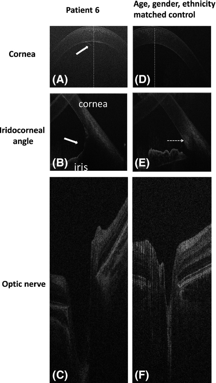
Spectral domain optical coherence tomography of case 6 with primary childhood glaucoma in the right eye (A–C) and the healthy control (D–F). The white arrow on figure A shows additional tissue on the endothelium in the centre of the cornea; arrow on figure B indicates abnormal iris insertion (iris root in front of the Schlemm canal) when compared to control; the horizontal tomogram of the optic nerve in the patient with glaucoma showed a larger horizontal diameter and depth of the cup as compared to the healthy control (F).