Figure 1.

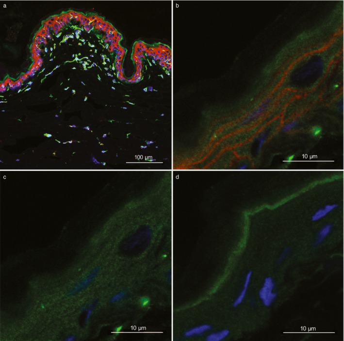
Frozen axilla skin, sections from German shepherd dogs with atopic dermatitis from one heterozygote (a, ID = 8) and one homozygote (b–d, ID = 1) for the top genome‐wide association study (GWAS) single nucleotide polymorphism (SNP) risk allele.1 (a, b) Immunostaining for PKP2 (green), plakoglobin (red) and 4′,6‐diamidino‐2‐phenylindole (DAPI) staining (blue) show both cytoplasmic and cell membrane expression of both PKP2 and plakoglobin in keratinocytes of the epidermis. In addition, PKP2 expression is present in other cell types in both epidermis and dermis. (c) The same picture as (b) with red and blue colours omitted, thus showing only PKP2 expression. (d) A negative control with only secondary antibodies showing nonspecific (green fluorescence) binding to stratum corneum. The protein expression of PKP2 in the keratinocyte cytoplasm is more homogenous compared with plakoglobin, while the expression at the cell border is more evident and distinct for plakoglobin. Magnification is ×20 (a), and ×63 with ×3.1 zoom (b–d). The scale bars are 100 μm (a) and 10 μm (b–d).
