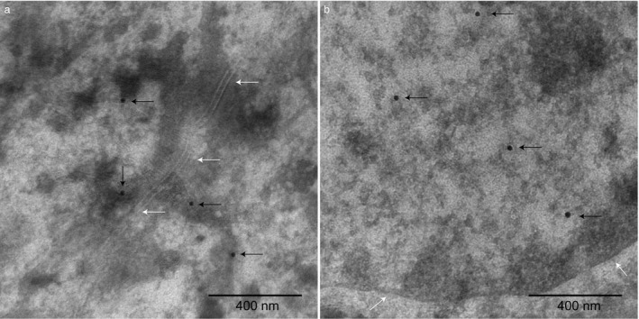
Figure 3.

Electron microscopy immunohistochemistry with gold particles (20 nm) indicate immunoreactivity to PKP2 in the epidermis in axilla skin from a control German shepherd dog (ID = 11) homozygous for the control allele at the top genome‐wide association study (GWAS) single nucleotide polymorphism (SNP).1 (a) Black arrows indicate gold particles binding to PKP2 in the vicinity of desmosomes on keratin filaments attached to desmosomes (desmosomes are marked with white arrows). (b) Black arrows indicate gold particles binding to PKP2 in the nucleus (the nucleus border is marked out with white arrows). The magnification is ×43,000 and scale bars 400 nm.
