
Figure 3.
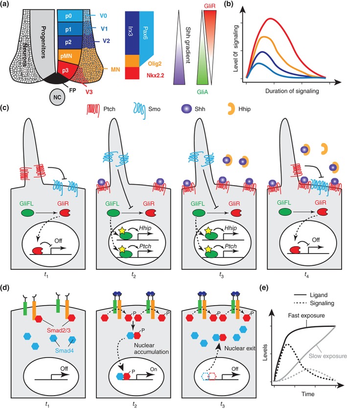
Pathway adaptation as means to measure signaling duration and dynamics. (a) The ventral neural tube is subdivided into different progenitor domains (p0–p3 and pMN) which form distinct classes of interneurons (V0–V3) and motor neurons. Subdivision of the three most ventral progenitor domains (p2, pMN, p3) is controlled by the differential expression of Irx3 (p2 only), Pax6 (p2, low in pMN), Olig2 (pMN), and Nkx2.2 (p3). This pattern is established in response to the graded activity of Shh, which generates two opposing gradients of activated Gli (GliA) and Gli repressor (GliR). (b) Due to pathway intrinsic negative feedback, different levels of Shh signaling are converted into distinct durations of Shh pathway activity. (c) Pathway intrinsic negative feedback leads to Shh pathway adaptation. In the absence of ligand (t 1), Ptch1 receptors block the entrance of Smo to the primary cilium. This leads to proteasomal degradation of full‐length Gli (GliFL) into its repressor version (GliR). Upon exposure to Hh ligands (t 2), Hh binds to Ptch1 and inhibits its activity, so Smo can enter into the cilium. Smo activity blocks Gli processing leading to stabilization and activation of GliFL, which activates target genes in the nucleus. Among the target genes of Gli are Hhip1 and Ptch1, which encode negative regulators of Hh pathway activity. Consequently, levels of Hh ligands need to rise continuously to inactivate increasing concentrations of Hhip1 and Ptch1 receptor (t 3). Failure to do so, results in sequestration of Hh ligands and inactivation of the pathway (t 4). (d) Pathway adaptation can be used to measure the speed of ligand exposure in the TGFβ pathway. In the absence of ligand, TGF receptors do not dimerize and Smad2/3 stay bound to the receptors (t 1). Upon ligand binding, receptors dimerize, and phosphorylate Smad2/3, which allows dissociation of phosphorylated Smad2/3 from the receptor, interaction with Smad4 in the cytoplasm, nuclear accumulation, and activation of target genes (t 2). Despite continuous exposure to the ligand, Smads are transported from the nucleus after a certain amount of time, leading to pathway adaptation (t 3). (e) Pathway adaptation can be used to measure the speed of ligand exposure. Fast exposure of ligand (solid black line) leads to high levels of signaling (dashed black line) before the pathway adapts. In contrast, the response to slower ligand exposure (gray lines) is dampened by pathway adaptation.