Figure 1.

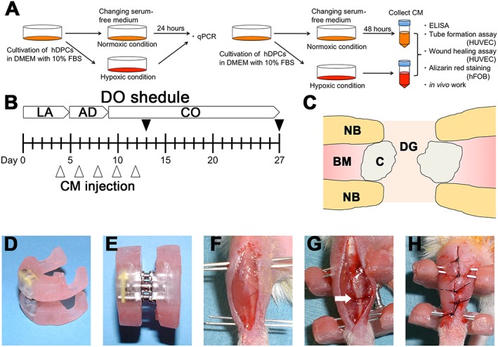
Outline of the study: (A) in vitro experiments and analyses; (B) in vivo distraction schedule and experimental design; after a 5 day latency period, distraction was started at a rate of 0.4 mm/12 h and continued for 4 days, increasing the length of the bone defect by 3.2 mm: black arrowheads, time points of sacrifice; white arrowheads, time points for injecting 20 µl of either control medium (control), CM–Nor or CM–Hyp; LA, latency period; AD, active distraction period; CO, consolidation period. (C) Schematic drawing of distraction site: NB, native bone; BM, bone marrow; C, callus; DG, distraction gap. (D–H) A custom‐made distractor was composed of two incomplete acrylic resin rings and an expansion screw: an anterior longitudinal incision was made on the right lower limb of each mouse; needles were inserted through the skin into the proximal and distal metaphysis of the tibia; subsequently, sets of needles were fixed to the custom‐made fixator with acrylic resin; after polymerization of the resin, osteotomy was carried out at the middle of the diaphysis (white arrow); the wound was closed with a 4–0 nylon suture. [Colour figure can be viewed at wileyonlinelibrary.com]
