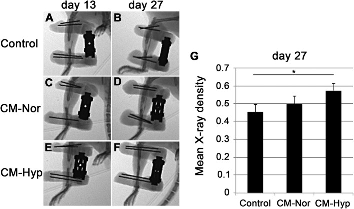
Figure 8.

CM from hypoxia promotes healing during DO: newly formed bone was assessed radiologically on days 13 (A, C, E) and 27 (B, D, F) (n = 5). There was no significant difference between CMs on day 13; on day 27 the controls (B) showed some calcified and narrow bone formation in the distraction site; the CM–Nor group showed calcified bone formation in the DO gap (D); the CM–Hyp group (F) had strongly calcified bone formation with high X‐ray density in the distraction site compared with controls (G). Data represent mean ± SD; *p < 0.05
