
Figure 4.
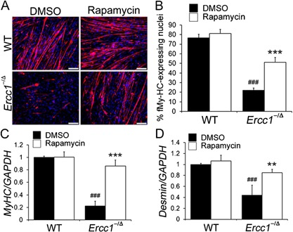
Measuring the effect of rapamycin on the myogenic differentiation of MDSPCs isolated from progeroid Ercc1 −/Δ mice. (A) Images of in vitro myogenic differentiation. Cells were immunostained for the terminal differentiation marker, f‐MyHC (red). Scale bar = 50 µm. (B) Quantification of myogenic differentiation was calculated as the fraction of cells (DAPI, blue) expressing f‐MyHC (red) from four independent MDSPC populations. Error bars indicate the standard deviation. Statistical significance was determined using one‐way ANOVA test with Tukey–Kramer post hoc test. ### p < 0.001 versus WT‐DMSO, ***p < 0.001 versus Ercc1 −/Δ‐DMSO. (C and D). Quantitative RT‐PCR to measure the expression levels of the myogenic differentiation markers MyHC and desmin after myogenic differentiation of MDSPCs isolated from Ercc1 −/Δ and WT mice, cultured with and without rapamycin. Error bars indicate the standard deviation. Statistical significance was determined using one‐way ANOVA test with Tukey–Kramer or Scheffe's post hoc test (n = 4). ### p < 0.001 versus WT‐DMSO, **p < 0.01, ***p < 0.001 versus Ercc1 −/Δ‐DMSO.