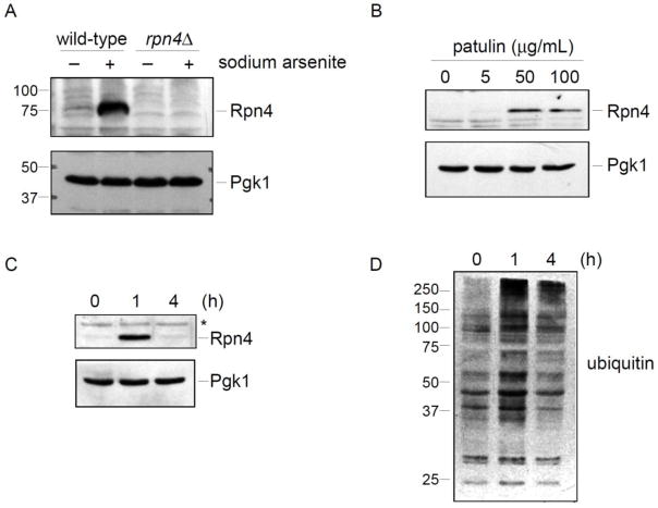
Figure 2. Patulin induces the Rpn4 stress response.
A) Validation of the Rpn4 antibody. Wild-type and rpn4Δ strains were treated with sodium arsenite (1 mM) for one hour. Whole cell extracts were prepared and evaluated by SDS-PAGE followed by immunoblot with anti-Rpn4 antibody (upper panel) or anti-Pgk1 antibody (lower panel; loading control).
B) Rpn4 protein levels after patulin treatment for one hour at the indicated concentrations. Whole cell extracts from a wild-type strain were prepared and analyzed by SDS-PAGE followed by immunoblot with anti-Rpn4 antibody (upper panel) or anti-Pgk1 antibody (lower panel; loading control).
C) Temporal dynamics of the Rpn4 response after induction by patulin (50 μg/mL). Whole cell extracts from a wild-type strain were prepared and analyzed by SDS-PAGE followed by immunoblot with anti-Rpn4 antibody (upper panel) or anti-Pgk1 antibody (lower panel; loading control). Asterisk, non-specific immunoreactive band.
D) Levels of ubiquitin conjugates in whole cell extracts, as determined by SDS-PAGE followed by immunoblot with anti-ubiquitin antibody. The same whole cell extracts were used to prepare the immunoblots in panels C and D. Therefore, the Pgk1 loading control of panel C applies to panel D as well.

