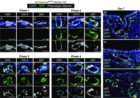
Figure 3.
ACTA2-CreERT2 lineage-traced myoepithelial cells express basal cell markers early during submucosal gland development. ACTA2-CreERT2:ROSA-TG mice were induced on the day of birth (0.2 mg) and harvested at 3, 5, 7, or 21 days of age. Tracheal sections for the four phases of gland development were immunostained for GFP, CK5, CK14, and/or p63. This procedure required antigen retrieval, which prevented the immunodetection of tdTomato. DAPI was used to mark nuclei. (A–C) Placode phase (phase 1), (D–F) elongation phase (phase 2), (G–I) branching phase (phase 3), and (J–L) differentiation phase (phase 4). Multichannel images are shown above single-channel images for each phase. (M–O) Low-power images of 7-day tracheal sections stained for each of the markers, as indicated. Scale bars: 25 μm.

