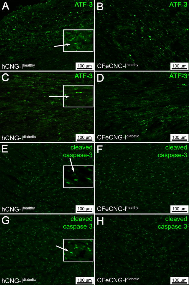
Fig. 1.

Distal nerve segments 56 days after delayed nerve reconstruction stained for ATF-3 and cleaved caspase-3. A–D Photomicrographs present immunohistology for ATF-3. E–H Photomicrographs present immunohistology for cleaved caspase-3. Inserts are magnifications from the image showing the oval ATF-3 (rabbit anti-ATF-3 polyclonal antibody) and cleaved caspase-3 (anti-cleaved caspase-3 antibody) stained cells interpreted as Schwann cells (secondary antibody Alexa Fluor 488 conjugated goat anti-rabbit IgG; green; see methods and Additional file 2: Figure 2). hCNG-I healthy hollow chitosan nerve guide from healthy rats, CFeCNG-I healthy chitosan film enhanced chitosan nerve guide from healthy rats, hCNG-I diabetic hollow chitosan nerve guide from diabetic GK rats, CFeCNG-I diabetic chitosan film enhanced chitosan nerve guide from diabetic rats. For details of the results of the quantification see Table 2. Scale bars display 100 µm in all images
