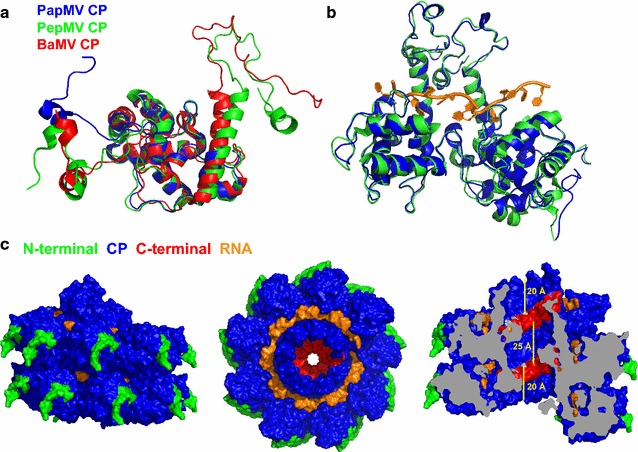
Fig. 1.

Modelling of the PapMV CP and PapMV structure. a The full-length structure of the PapMV CP was modelled based on the recently published structure of two members of the potexvirus group: BaMV CP and PepMV CP. The core region of PapMV CP (PDB 4DOX, blue) superimposes well on the core region of PepMV CP (PDB 5FN1, green) and BaMV CP (PDB 5A2T, red). b Superposition of two subunits of the PapMV model (blue) with two CP subunits of the PepMV structure (green, PDB 5FN1) demonstrates the concordance between the two structures. c To show how each of the PapMV CP interacts with each other, and with the ssRNA in the nanoparticle, we modeled the self assembly of the 18 subunits (~2 turns) that comprise PapMV nanoparticles. The CP N-terminal residues (10 first) are shown in green, the CP core in blue, the CP C-terminus in red (10 last residues), and the RNA is in orange. The last C-terminal residue of the CP is displayed in light grey in the cutaway view on the right, and the bars represent 20 Å—the distance separating CP C-terminal residues from the PapMV nanoparticle exterior at both extremities, and 32 Å—the distance between two CP C-terminal residues separated by one capsid turn
