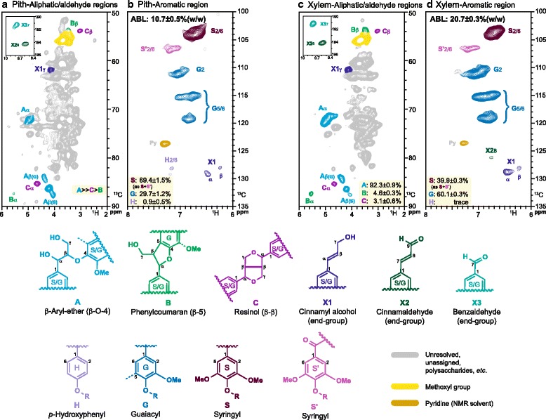
Fig. 1.

2D–HSQC-NMR spectra of pith and xylem tissues. a, c Aliphatic/aldehyde and b, d aromatic regions of 1H–13C (HSQC) spectra from cell wall gel samples in DMSO-d6:pyridine-d5 (4:1). a, b: Pith tissue. c, d: Xylem tissue. Levels of A, B and C in pith (A) were not quantified because of co-located polysaccharides with Aα, however the spectra allow the relative estimation. NMR lignin correlations are coloured to match the structures responsible. ABL refers to total lignin content measured as acetyl bromide lignin
