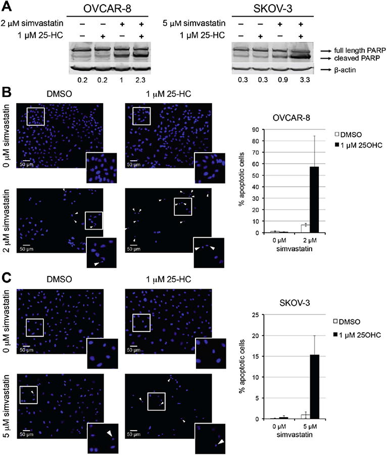
Fig. 2.

25-hydroxycholesterol combined with simvastatin increases apoptosis in ovarian cancer cell lines. A) Immunoblots of PARP after 48 h of treatment with indicated concentrations of statins and 25-HC. Band quantification represents the fraction of cleaved PARP after normalization to β-actin. B and C) DAPI stained cells after 72 h of treatment as indicated. The cells were scored as either apoptotic or non-apoptotic based on nuclear morphology. Representative images of OVCAR-8. (B) and SKOV-3 (C) are shown. Arrows indicate apoptotic cells and magnification of cells in the white squares are shown in the lower right corner. Percentages of apoptotic cells relative to vehicle are graphed.
