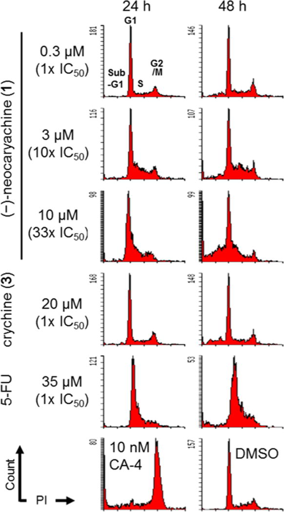
Figure 1.
Effects of (−)-neocaryachine (1) and crychine (3) on cell cycle progression in MDR cells. KBKB-VIN cells were treated with (−)-neocaryachine (1), crychine (3), or 5-FU for 24 or 48 h or CA-4 for 24 h at the indicated concentrations. DMSO was used as a vehicle control. Cell cycle distributions of treated cells were analyzed by flow cytometry after staining with propidium iodide (PI).

