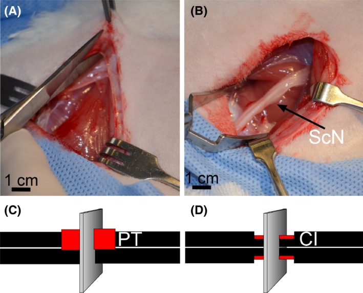
Figure 1.

An experimental model of nerve injury. (A,B) Accessing the sciatic nerve of a rabbit. (C) PT nerve injury in which 50% of the thickness of the nerve for a length of 1 cm is removed. (D) CI nerve injury in which a hemostat is placed around the sciatic nerve 1 cm distal to the sciatic notch. The hemostat is then tightened to the first locking flange and held in place for 30 s. In panels C and D, the sites of injury are indicated by red color. Planes corresponding to the orientation of samples collected from the injured nerves are also indicated
