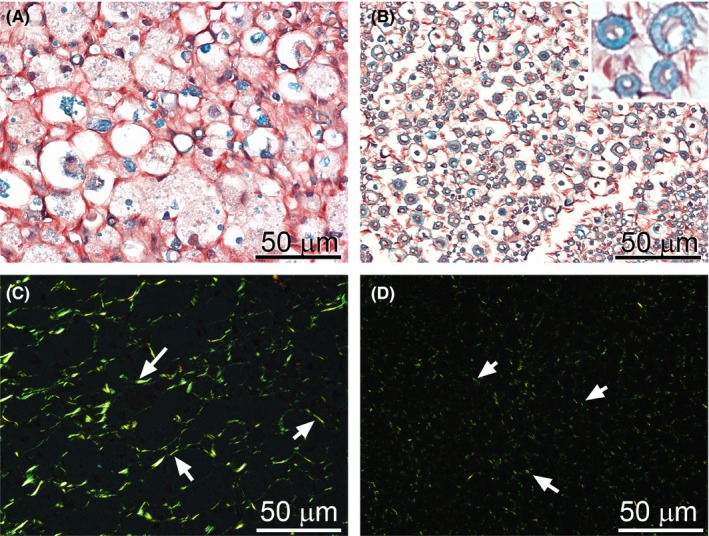
Figure 3.

Morphology of the CI nerves stained with luxol fast blue for myelin and with Sirius for collagen deposits. In a crushed nerve (A), degradation of myelin is clearly apparent; in uninjured nerve, abundant myelinated axons are present (B, insert). Observation of Sirius‐stained samples in a polarizing microscope demonstrated an increase in collagen fibrils in the endoneurium of injured nerves (C, arrows). In contrast, in the control, collagen fibrils were sparsely distributed throughout endoneurial space (D, arrows)
