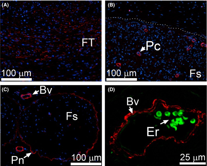
Figure 6.

αSMA immunostaining of the CI nerves. (A) αSMA‐positive cells are evident in perineural FT. (B) Within fascicles (delineated with a dotted line) αSMA‐positive staining is only seen in pericytes (Pc). (C) In uninjured control αSMA‐positive staining is also observed around endoneurial blood vessels (Bv) and within perineurium (Pn). (D) A high magnification of a blood vessel identified by the presence of erythrocytes (Er) seen due to green autofluorescence
