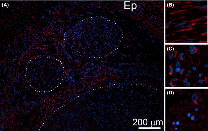
Figure 7.

HSP47‐positive cells are evident within the CI nerves. (A) A representative region depicting fascicles (delineated with dotted lines) and epineurium (Ep). (B) A magnified view at HSP47‐positive cells present in epineurium of a CI site. (C) A magnified view at HSP47‐positive cells present within endoneurium of a CI site. (D) A magnified view at HSP47‐positive cells present within endoneurium of a control uninjured nerve
