
Figure 3.
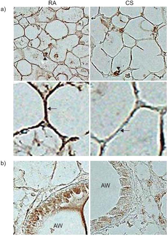
Reduced leptin receptor expression in lung parenchyma of AKR/J mice exposed to 4 months of cigarette smoke (CS) or room air. Representative images of a minimum of three mice per condition. a) Decreased immunohistochemical staining of leptin receptor (brown) in alveolar walls of smoke-exposed mice using polyclonal antibody against ObR and avidin-biotin-peroxidase complex method. Arrowheads denote preserved leptin receptor staining of alveolar macrophages after CS exposure. Arrows show reduced airspace wall staining in smoke-exposed mice. b) Reduced leptin receptor expression is also evident in airway epithelial cells of smoke-exposed mice. RA: room air exposed; AW: airway lumen.