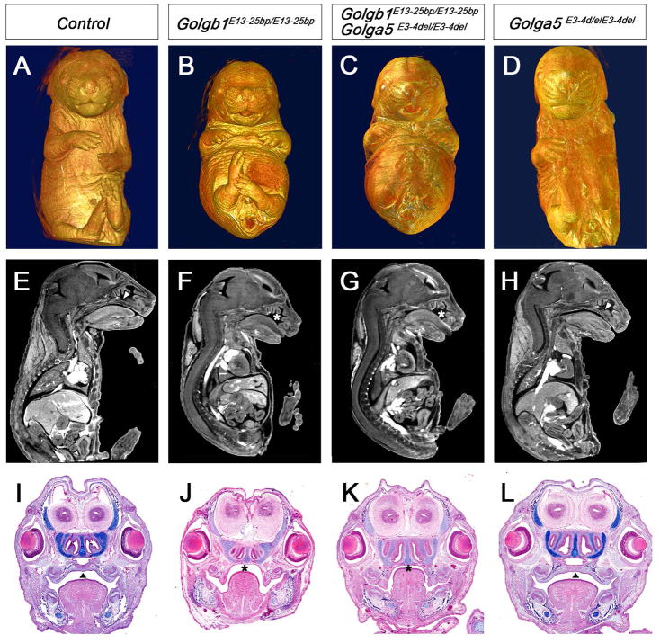
Figure 5. Analysis of Golga5/Golgb1 double mutant mice.
(A–D) Whole mount ventral views of Micro-CT scanned and 3D reconstructed E17.5 control (A), Golgb1E13-25bp/E13-25bp homozygous (B), Golga5E3-4del/E3-4delGolgb1E13-25bp/E13-25bp double homozygous (C), and Golga5E3-4del/E3-4del mutant (D) embryos. (E – H) Representative mid-sagittal Micro-CT scans of E17.5 control (E), Golgb1E13-25bp/E13-25bp homozygous (F), Golga5E3-4del/E3-4delGolgb1E13-25bp/E13-25bp double homozygous (G), and Golga5E3-4del/E3-4del mutant (H) embryos. (I–J) Coronal histological sections of E16.5 embryos showing control (I), Golgb1E13-25bp/E13-25bp mutant (J), Golga5E3-4del/E3-4delGolgb1E13-25bp/E13-25bp double mutant (K), and Golga5E3-4del/E3-4del mutant (L) embryos. Arrowheads in E, H, I, and L point to normal fused secondary palate. Asterisk in F, G, J, and K marks the cleft palate defect.

